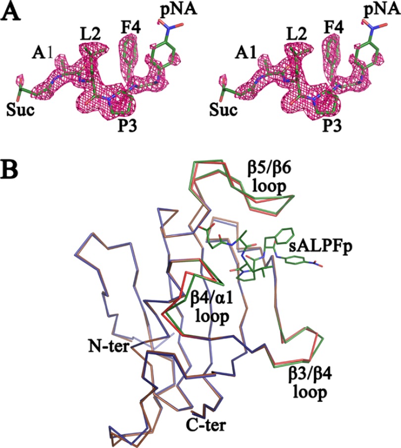
Fig 3.
Stereoview of sALPFp peptide and overlay of apo PvFKBD35 and sALPFp-bound PvFKBD35. (A) Stereoview of the electron density map (2FO-FC) contoured at the 1.0 σ level for the peptide molecule. It could be clearly seen that the Leu-Pro peptide bond adopts a cis-isomer conformation. (B) Superposition of the Cα traces of apo PvFKBD35 colored in blue and PvFKBD35 bound to sALPFp colored in brown. The ligand-flanking loops of the apo- and sALPFp-bound structures are colored in red and green, respectively. The sALPFp peptide is shown as green sticks for reference.

