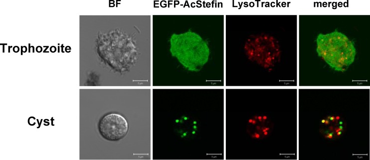
Fig 2.
Intracellular localization of AcStefin protein. Shown are subcellular localization changes in EGFP-fused AcStefin. Trophozoites were transfected with pUb-EGFP-AcStefin (green), transferred into a medium to induce encystation, incubated for 72 h, and examined under a fluorescence microscope. Lysosomes and autophagolysosomes were visualized by LysoTracker Red staining (red), and the resulting merged images are shown (yellow). BF denotes bright-field images. Bar = 5 μm.

