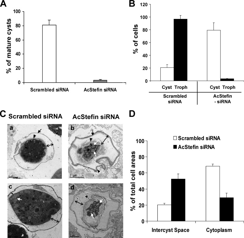
Fig 4.
Effects of AcStefin knockdown on encystation and excystation. (A) The same numbers of mature cyst-like cells with a cyst wall of scrambled siRNA transfectants (control) (light bar) and AcStefin-siRNA transfectants (dark bar) were collected under a microscope at 72 h postencystation and then treated with 0.05% Sarkosyl for 30 min. Sarkosyl-resistant mature cysts were stained with 0.22% trypan blue and counted under a microscope. (B) The same numbers of mature cyst-like cells were transferred onto E. coli-covered nonnutrient agar plates and incubated for 7 days at 27°C, and trophozoites (light bars) and cysts (dark bars) were counted for control (scrambled-siRNA-transfected) and AcStefin-siRNA-transfected (AcStefin-siRNA) cells. Value represent the means ± standard deviations of triplicate experiments. (C) Representative TEM images showing a thick intercyst space and reductions in the cytoplasmic areas of AcStefin knockdown cells. Cells transfected with nonspecific scrambled siRNA (a and c) and AcStefin siRNA (b and d) were transferred into encystment medium, incubated for 72 h, and examined by TEM. The black arrows and arrowheads show the exocyst wall and the endocyst wall in both scrambled-siRNA- and AcStefin-siRNA-transfected cells, respectively. The black double-headed arrows indicate the intercyst space. The white arrows indicate lipid droplets. Bar = 2 μm. (D) Intercyst space and cytoplasmic areas were measured. Pixel areas were determined by using ImageJ analysis tools, using 30 cysts of scrambled-siRNA- or AcStefin-siRNA-transfected cells in each of 10 TEM pictures.

