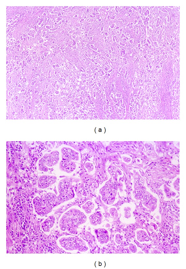
Figure 3.

Histological examination of the specimen revealed that the tumor consisted of an invasive micropapillary component. Carcinoma cell clusters were floating in the clear spaces. (a) Hematoxylin-eosin stain, original magnification, ×40. (b) Hematoxylin-eosin stain, ×200.
