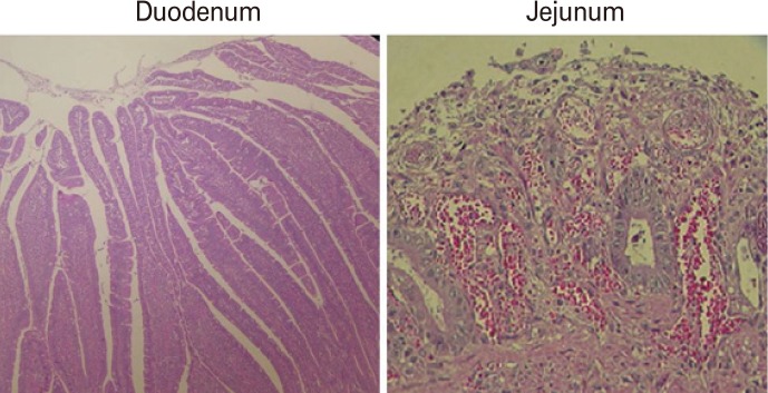
Fig. 6.
Histopathological changes in the small intestine of dogs in group II. Hematoxylin and eosin staining was performed on the samples from duodenum and jejunum of the group II dogs that showed typical clinical signs of canine parvovirus infection. Histopathological changes in 2 dogs (dogs #3 and #4) were similar, and representative changes are shown here.

