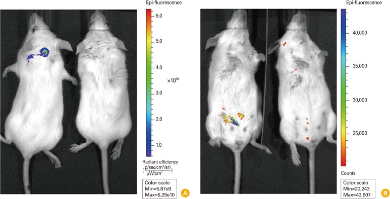
Fig. 2.
Visualization of E. coli-GFP strain in mice by in vivo imaging. (A) Fluorescent imaging of E. coli-GFP injected with intradermal route. 5×10⁹ CFU (500 µL) of E. coli-GFP was subcutaneously injected and dorsal side of mice were analyzed. Left animal, injected mouse; right animal, uninjected mouse; pseudo-color, red (high) to blue (low). (B) Fluorescent imaging of E. coli-GFP injected with intraperitoneal route. 3.5×10⁹ CFU (350 µL) of E. coli-GFP was injected to abdominal cavity and ventral side of injected mice were analyzed. Left animal, injected mouse; right animal, uninjected mouse; pseudo-color, blue (high) to red (low). E. coli-GFP, Escherichia coli MC1061 strain which expresses green fluorescent protein; CFU, colony-forming unit.

