FIGURE 6:
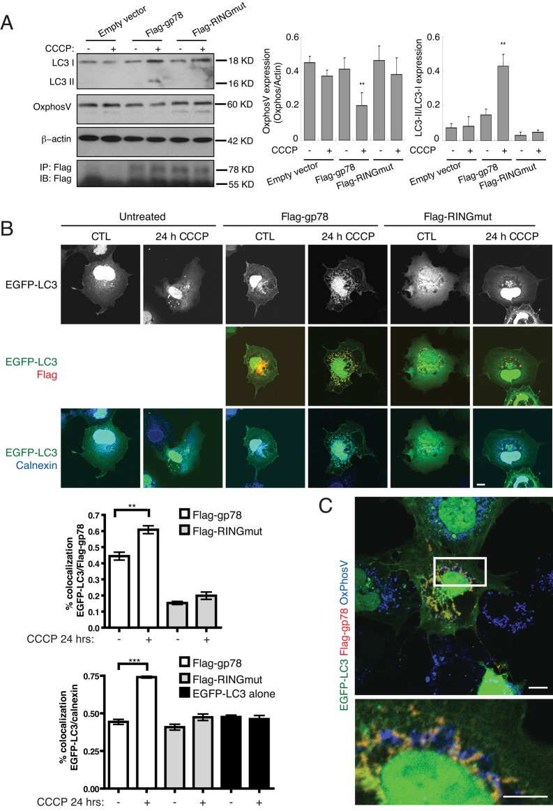
Gp78 recruits the autophagosomal marker LC3 to the ER at sites of mitochondrial degradation. (A) Western blots show expression of LC3-I and LC3-II isoforms and of mitochondrial OxPhosV in Cos7 cells transfected with pcDNA (empty vector), Flag-gp78, or Flag-RINGmut and treated or not with 10 μM CCCP for 2 h. OxPhosV expression was quantified by densitometry relative to β-actin and LC3-II relative to LC3-I (n = 3, ±SEM; *p < 0.05, **p < 0.01). (B) Cos7 cells were transfected with EGFP-LC3 alone or cotransfected with EGFP-LC3 and either Flag-gp78 or Flag-RINGmut and treated with 10 μM CCCP for 24 h. Paraformaldehyde (PFA)-fixed cells were labeled with anti-Flag (red) and anti-calnexin (blue) antibodies and EGFP-LC3 fluorescence (black and white; green) imaged directly. Nuclear EGFP-LC3 label is saturated to highlight cytoplasmic and ER-localized EGFP-LC3. Bar graphs show colocalization of EGFP-LC3 and either Flag-gp78 or calnexin (mean ± SEM, n = 3, ±SEM; **p < 0.01; ***p < 0.005; bar, 10 μm). (C) Cos7 cells cotransfected with EGFP-LC3 and Flag-gp78 were treated with CCCP for 24 h and after PFA fixation labeled with anti-Flag (green) and anti-OxPhosV (blue) antibodies. EGFP-LC3 fluorescence (green) was imaged directly. Residual mitochondria in Flag-gp78 transfected cells are closely associated with the EGFP-LC3–labeled ER. Bar, 10 μm.

