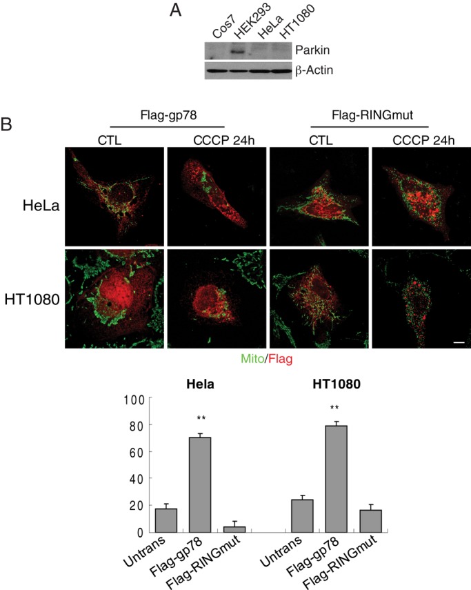
FIGURE 7:

Gp78 promotes CCCP-induced mitophagy in Parkin-null HeLa cells. (A) Cos7, HeLa, HEK293, and HT-1080 cells were western blotted for Parkin and β-actin. (B) Parkin-deficient HeLa and HT-1080 cells were transfected with Flag-gp78 or Flag-RINGmut and either untreated (CTL) or treated with CCCP for 24 h, as indicated, and labeled with antibodies to Flag and OxPhosV (mito). Bar graphs show the percentage loss of mitochondrial mass of CCCP-treated cells relative to untreated cells ((mitochondrial area[CTL − CCCP]/mitochondrial area[CTL]) × 100). Mean ± SEM; 10–20 cells/experiment; n = 3, **p < 0.01; bar, 10 μM; see Supplemental Table S1 for mitochondrial area values.
