Figure 1.

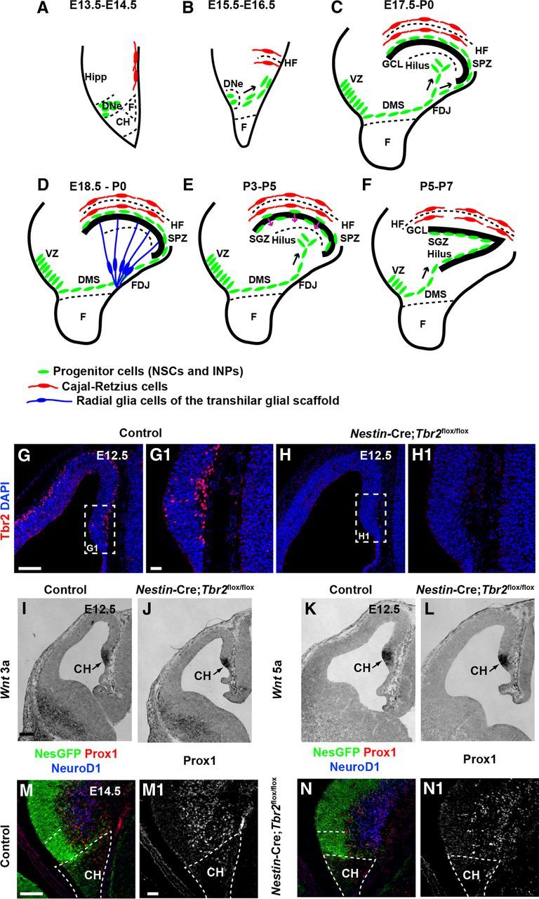
Early development of the DG is essentially normal in Nestin-Cre;Tbr2flox/flox mice. A–F, Schematic diagram illustrating the steps in DG development. Green represents progenitor cells (NSCs and INPs); red, Cajal-Retzius cells; blue, transhilar radial glial scaffold. A, B, Progenitor cells initially located in the DNe migrate to the primordial DG concurrent with invagination of the pial surface and migration of Cajal-Retzius cells to the HF between E13.5 and E16.5. C, Continued migration of progenitors through the DMS contributes to formation of the SPZ during later stages of development (E17.5-P0). D, The transhilar radial scaffold forms at approximately the same time as radial glia become localized to the FDJ. E, Transition of progenitor cells out of the SPZ occurs between P3-P5. F, The SGZ neurogenic niche is established by P5-P7. G, G1, Tbr2 protein (red) is expressed in cells in the cortical hem (CH) at E12.5 in control mice. H, H1, Tbr2 protein is ablated in the cortical hem of mutant mice by E12.5. White dashed boxes in G and H represent areas shown at higher magnification in G1 and H1, respectively. I–L, Expression of Wnt3a and Wnt5a, markers of the cortical hem, are present at approximately normal levels in Nestin-Cre;Tbr2flox/flox mice. M, N, M1, N1, Markers of DG granule neurons (NeuroD1, Prox1) are present in the primordial DG of mutant mice but are slightly reduced compared with controls as early as E14.5. Scale bars: G, 100 μm; G1, 20 μm; I, 100 μm; M, 75 μm; M1, 15 μm.
