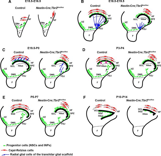
Figure 9.
Schematic diagram summarizing DG defects in Nestin-Cre;Tbr2flox/flox mice. A, Initial invagination of the pial surface is delayed in Nestin-Cre;Tbr2flox/flox mice, and the migration of Cajal-Retzius cells (red) to the forming HF is delayed in mutants. B, Development of the transhilar radial glial scaffold (blue cells) is abnormal in Nestin-Cre;Tbr2flox/flox mice by E18.5-E19.5, and the number of Cajal-Retzius cells (red) populating the HF is reduced. C, Blbp+ radial glial cells (blue) that contribute to the transhilar radial glial scaffold complete their redistribution to the HF by E19.5-P0 in control mice, but this migration is delayed in Nestin-Cre;Tbr2flox/flox mice. D, During early postnatal development (P3-P4), progenitor cells (green) populating the transient SPZ are redistributed to form the SGZ niche. In Nestin-Cre;Tbr2flox/flox mice, these cells are retained in the SPZ and fail to migrate to the SGZ. E, Formation of the SGZ (green, progenitor cells) is complete by P5-P7 in control mice, and the SPZ is no longer apparent. In mutant mice, retention of progenitors in the SPZ persists. F, By P10-P14, both the suprapyramidal and infrapyramidal blades of the DG are formed in control mice, and progenitor cells (NSCs and INPs, green) are localized to the SGZ. In Nestin-Cre;Tbr2flox/flox mice, the infrapyramidal blade fails to form, the suprapyramidal blade is reduced in size, and progenitor cells are nearly absent from the SGZ (green).

