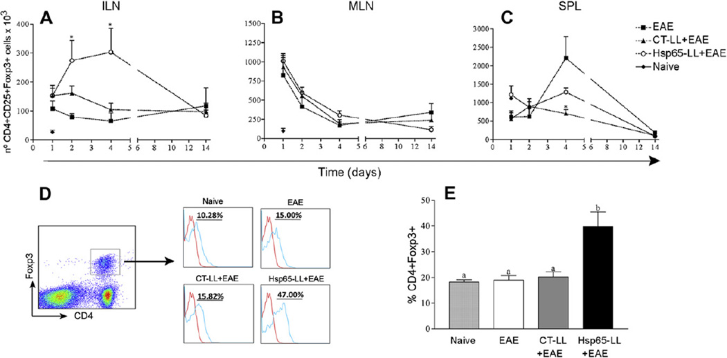
Fig. 4.
Oral pretreatment of mice with M. leprae-Hsp65-producing L. lactis lead to an increase in both natural and inducible CD4+Foxp3+ Treg cell populations in mice. C57BL/6 Foxp3-GFP Knock-in mice were fed or not (Naïve) medium (EAE), wild type (CT-LL+EAE) or M. leprae-Hsp65-producing L. lactis (Hsp65-LL+EAE) for four days and EAE was induced ten days later. After 1, 2, 4 and 14 days, mice were killed and inguinal (ILN; A) and mesenteric lymph nodes (MLN; B) and spleen (SPL; C) removed. Cells were stained with Cy-anti-CD4 and PE-anti-CD25. CD4+ cells were gated. Bar graphs are shown as mean ± SEM. ANOVA, post-test Tukey. *Statistically different from EAE group; p < 0.05. (D and E) Increase in the natural regulatory T (nTreg) cells population in inguinal lymph node after oral administration of M. leprae-Hsp65-producing L. lactis. C57BL/6 Foxp3-GFP Knock-in mice were fed or not (Naïve) medium (EAE), control (CT-LL+EAE) or M. leprae-Hsp65-producing L. lactis (Hsp65-LL+EAE) for four days and EAE was induced ten days later. Four days later, mice were killed and inguinal lymph nodes removed. Cells were stained with Cy-anti-CD4 and PE-anti-Helios. CD4+Foxp3+ cells were gated. Plots are representative of the mean of 4 mice/group and data are representative of three independent experiments. Bar graphs are shown as mean ± SEM. ANOVA, post-test Tukey. *Statistically different from EAE group; p < 0.05.

