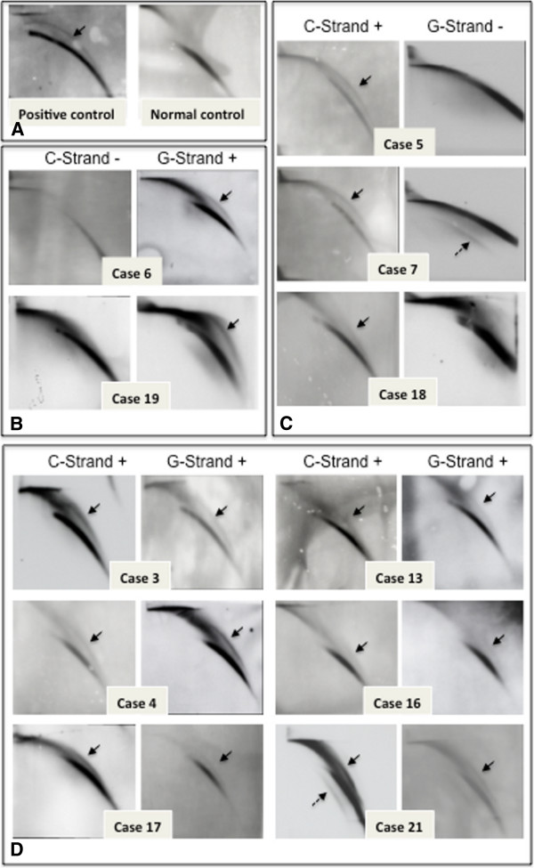
Figure 2.

Blots from 2D gels of telomeres showing the presence of ECTR molecules in CML samples. (A) The presence and absence of the circular ECTR detected in C strand in a positive (left) and a negative (right) control sample. (B) Samples with circular ECTR in G-strand. (C) Samples with circular ECTR in C-strand. (D) Samples with circular ECTR in both C- and G-strands. Note that case 7 and case 21 also have linear ECTR molecules (dashed arrows).
