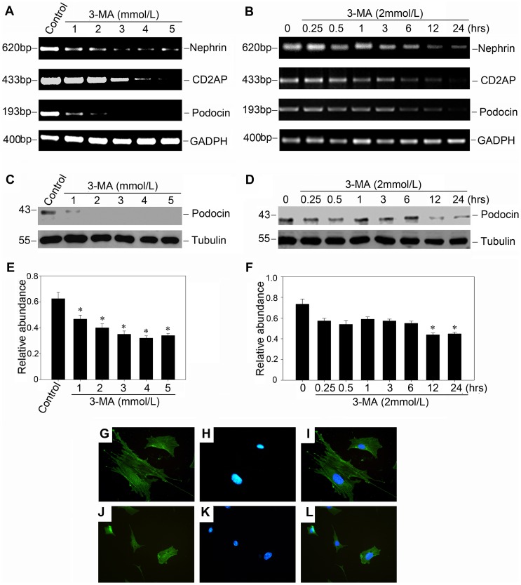
Figure 3. Autophagy inhibition by 3-MA suppresses the protein and mRNA expression of podocyte slit diaphragm proteins in a dose-dependent and time-dependent manner.
(A) and (B): RT-PCR demonstrates that 3-MA (2 mmol/L) inhibited the mRNA expression of podocyte slit diaphragm proteins such as nephrin, CD2AP and podocin in a dose-dependent and time-dependent manner. Podocytes were incubated with either increasing amounts of 3-MA for 24 hours (A), or the same concentration of 3-MA (2 mmol/L) for various periods of time as indicated (B). (C) and (D): Western blot analysis shows that 3-MA (2 mmol/L) inhibited podocin protein expression in a dose- and time-dependent manner. Cell lysates were immunoblotted with Ab's against podocin and α-tubulin, respectively. (E) and (F): Quantitative determination of podocin protein abundance after normalization with α-tubulin. Data are presented as mean ± SEM of three independent experiments. *P<0.05 vs. normal control; (G–L): Immunofluorescence staining shows the localization of podocin in the control and 3-MA treated podocytes. (G–I): control group; (J–L): podocytes incubated with 3-MA (2 mmol/L) for 24 hours.

