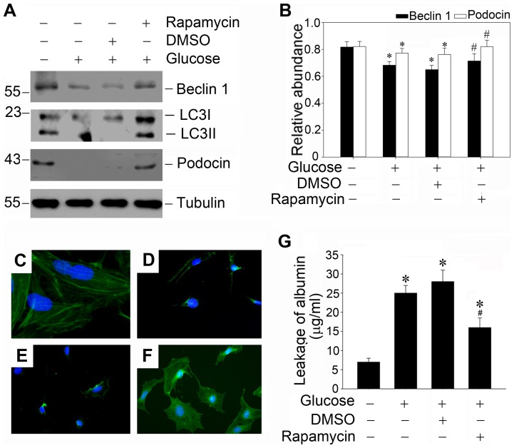
Figure 7. Rapamycin restores defective autophagy induced by high glucose and protects against high glucose induced podocyte injury.
(A) Western blot analysis shows that low concentration of rapamycin (1 ng/ml) could restore defective autophagy induced by high glucose and protect against podocyte injury. Podocytes were pretreated with DMSO or 1 ng/ml rapamycin for 0.5 h, and followed by incubation with high glucose (25 mmol/L) for 48 hours. Whole-cell lysates were immunoblotted with specific antibodies against Beclin-1, LC3, podocin or α-tubulin, respectively. (B): Quantitative determination of Beclin-1 protein and podocin protein abundance after normalization with α-tubulin. Data are presented as mean ± SEM of three independent experiments. *P<0.05 vs. normal control; # P<0.05 vs. high-glucose-treated group. (C–F): Representative photographs of podocin visualized by indirect immunofluorescence staining in the control and treated podocytes as indicated. (C): control group; (D): high-glucose-treated group; (E): DMSO pretreated + high glucose treated group; (F): rapamycin pretreated + high glucose treated group; (G): Graphic presentation of the albumin influx across podocyte monolayer. Podocyte monolayer on collagen-coated transwell filters was incubated with various treatments as indicated for 48 hours. Data are presented as mean ± SEM of three independent experiments. * P<0.05 vs. normal control; # P<0.05 vs. high-glucose-treated group.

