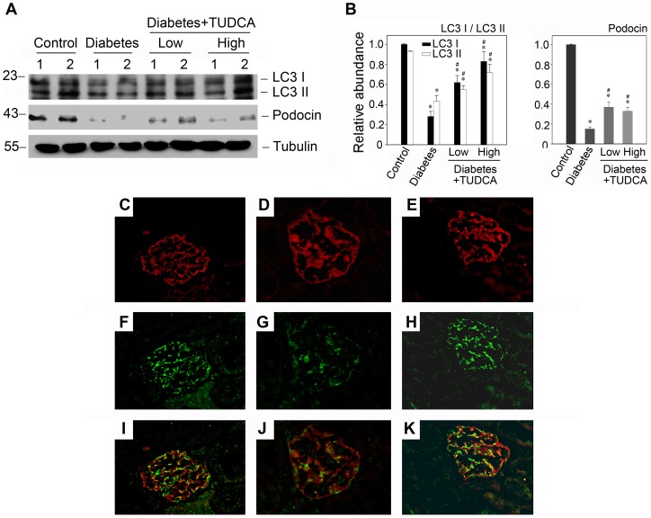
Figure 10. TUDCA restores the suppressed autophagy in diabetic mice and attenuates podocyte injury.
(A) Western blot analysis demonstrates an improvement of autophagy level and podocin expression in the glomeruli isolated from mice as indicated. The glomerular lysates (made from the pool of kidneys from six animals/group) were separated on a SDS-polyacrylamide gel and immunoblotted with a specific monoclonal antibody against LC3, podocin and α-tubulin, respectively. Samples from two individual animals were used at each timepoint. (B) Quantitative determination of LC3 and podocin protein abundance after normalization with α-tubulin. Data are presented as means ± SEM of three experiments. n = 6, *P<0.05 vs. normal control. #P<0.05 vs. diabetic group. (C–K) Immunofluorescence staining shows the changes of autophagosomes in various groups (400× magnification). Kidney section were immunostained with anti-LC3 antibody (green) to identify autophagosomes, followed by staining with anti-podocin antibody (red) to sever as a marker for podocytes. The left column (C, F and I): control group; The second column (D, G and J): diabetic group; The third column (E, H and K): diabetic group treated with 500 mg/kg/day TUDCA.

