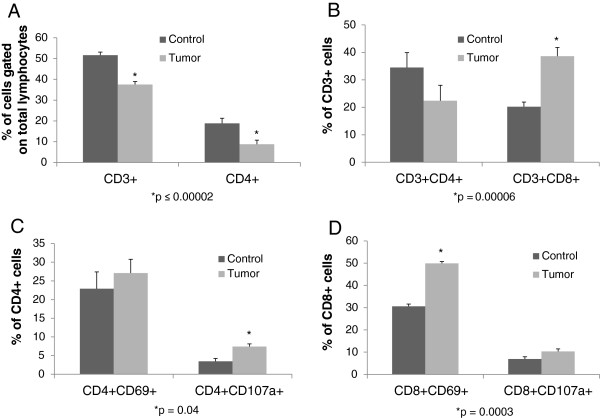
Figure 2.
Phenotype and function of T-cell subsets in colorectal liver metastases: Intrahepatic lymphocytes and tumor infiltrating lymphocytes were isolated. Lymphocyte populations were decreased in the metastatic deposits compared to lymphocytes isolated from livers harvested from control animals, though the percentage of CD3 + CD8+ T-cells was increased, and overall reflected an activated phenotype. A. CD3+ cells as a percentage of total lymphocytes in metastatic tumor tissue was lower compared to the number of CD3+ T cells in corresponding control liver (mean ± SEM : 37.5% ± 1.4 vs. 51.6 % ± 1.5, p = 0.00001, n = 11 per group). CD4+ cells as a percentage of total lymphocytes in metastatic tumor tissue was lower compared to the number of CD4+ T cells in corresponding control liver (8.7% ± 1.9 vs. 18.8% ± 2.5, p = 0.00002, n = 11 per group). B. CD3 + CD4+ populations trended to be less in tumor deposits than normal liver (22.4% ± 5.6 vs. 34.5% ± 5.4, p = 0.14, n = 11 per group). CD3 + CD8+ cells were significantly increased in the CRLM (38.6% ± 3.2 vs. 20.2% ± 1.7, p = 0.00006, n = 11 per group). C,D. CD4 + CD107a + and CD8 + CD69+ populations were increased in CRLM (7.4% ± 0.7 vs. 3.5% ± 0.8, p = 0.04 ; 50% ± 0.9 vs. 30.6% ± 0.5 p = 0.0003, respectively, n = 3 per group) . There was no difference in CD4 + CD69+ cells between the groups and a trend towards increased CD8 + CD107a + expression on TIL compared to non-TIL (27.1% ± 3.6 vs. 22.9% ± 4.5, p = NS ; 10.3% ± 1.2 vs. 6.9% ± 0.6, p = 0.08 , respectively, n = 3 per group). CRLM = colorectal liver cancer metastases. TIL = tumor infiltrating lymphocytes.

