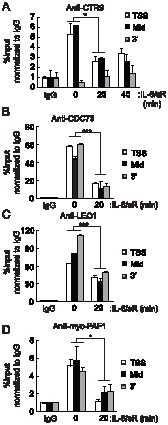
Figure 4. PAFc dissociates from the c-Fos locus after IL-6 stimulation. HepG2 cells were treated with IL-6 plus IL-6sR (20 ng/ml each) for the indicated length of time.

Soluble chromatins were immunoprecipitated with anti-CTR9 antibodies, and the bound DNAs were analyzed by quantitative PCR using primers specific to c-Fos (A). (B,C) CDC73 and LEO1 associations with c-Fos locus were analyzed after IL-6 plus IL-6sR (20 ng/ml each) stimulation. (D) Cells were transfected with Myc-PAF1 and 48 hours later, cells were treated with IL-6 plus IL-6sR (20 ng/ml each) for 20 minutes. ChIP assay was performed with anti-Myc antibody and bound DNAs were analyzed by quantitative PCR using primers specific to c-Fos. *p<0.05, ***p<0.001 by Student's t test. Error bars represents SD (n = 3).
