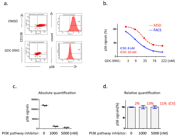
Figure 2.
Development of phospho flow assay. (a). PD response revealed by phospho flow analysis in MM1s cells. Note the peak shift of pS6 on the histogram in the GDC-0941-treated MM1s cells compared to DMSO. Baseline is defined based on isotype staining. (b). Cross-platform comparison between MSD and phospho flow cytometry. iMFI methodology is used to calculate the pS6 signals (c and d). Intra-assay variation studies demonstrate the absolute quantification of pS6 signals (c) and relative quantification (d), highlighting the coefficient of variation (CV). Note that MM1s cells were treated with DMSO or a PI3K pathway inhibitor (tool compound) for 2 hours in the above studies. Data shown (a-d) are representative of at least three independent studies.

