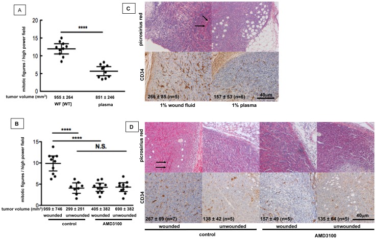
Figure 3. Wound-induced SDF-1α/CXCR4 signaling in tumor cells alters tumor cell proliferation, stromal composition and vascularization of tumors.
A, C. BALB/c mice were inoculated with 4T1 cells that were pre-treated for 5d with wound fluid or mouse plasma. B, D. BALB/c mice were inoculated with 4T1 cells and underwent wounding or sham surgery 9 days later. CXCR4 signaling was systemically inhibited by AMD3100. A, B. Mitotic figures in tumor sections. A. Unpaired t-test, p<0.0001, n = 10 specimens/group, observation time: 18 days. B. Bonferroni’s Multiple Comparison test, p<0.0001, n = 8–12 specimens/group, observation time: 28, mean ±95% CI. C, D. Top: Collagen staining with Picrosirius red. Bottom: CD34-positive blood vessels in tumors. Numbers in the lower left corner of images represent the density of CD34-positive structures/mm3. C. p = 0.0173, Mann-Whitney test. D. p = 0.0155, ANOVA, mean ±95% CI).

