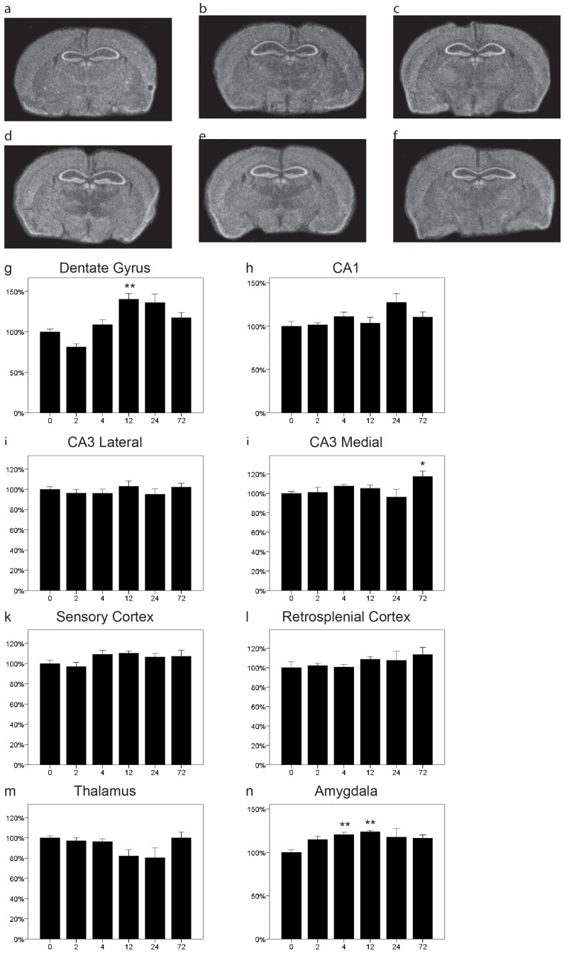
Figure 5. Distribution and regulation of Nogo-A mRNA in the mouse brain.
Representative coronal sections of control (a) and kainic acid injected (b 2 h, c 4 h, d 12 h, e 24 h, f 72 h) mice probed for Lotus mRNA by quantitative in situ hybridization. The expression of Nogo-A mRNA was stable in most regions of the brain, including CA1 (h), lateral (i) and medial (j) CA3, sensory (k) and retrosplenial cortex (l) as well as thalamus (m). However, in the dentate gyrus (g) and the amygdala (n) expression of Nogo-A transcripts was significantly increased with a peak at 12 h (i). Error bars represent S.E.M and all expression values were normalized to expression in control mice (t = 0). *p<0.05, **p<0.01 compared to 0 h.

