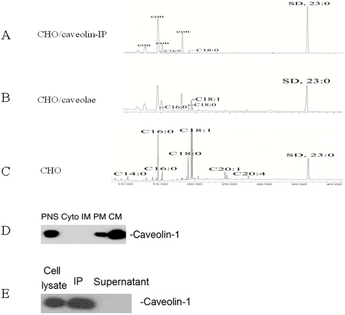
Figure 2. Quantification of fatty acids bound to caveolin-1, associated with caveolae, and present in CHO cells.
The CHO cells were cultured in Ham’s F-12 medium to 90% confluency. After washed with PBS, they were dissolved in MBST/OG on ice. Post nuclear supernatant (PNS), cytosol (Cyto), internal membranes (IM), plasma membrane (PM), and caveolae (CM) were isolated with Opti-Prep method. Total fatty acids were extracted from each sample with Folch reagent, methyl esterified with BF3, and then subjected to GC/MS equipped with Omegawax 250 capillary column. Fatty acids bound to caveolin-1 (A), associated with caveolae (B), and present in CHO cells (C) were identified by MS. (D) Isolation of caveolae from CHO cells. Subcellular fractions were isolated with Opti-Prep method and subjected to Western blot using antibody against caveolin-1. (E) Immunoprecipitation of caveolin-1. The CHO cell lysates were immunoprecipitated with anti-caveolin-1 IgG/protein A-Sepharose beads and detected by Western blot using antibody against caveolin-1. The experiments were repeated three times with triplicate measurements. Quantitative analysis of the data is shown in Figure 3.

