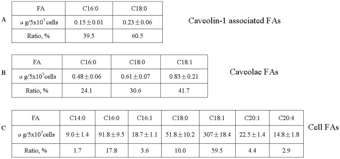
Figure 3. Quantification of fatty acids bound to caveolin-1, associated with caveolae, and present in CHO cells.
The CHO cells were cultured in Ham’s F-12 medium to 90% confluency. Caveolae were isolated with Opti-Prep method and caveolin-1 was purified by immunoprecipitation. Total fatty acids were extracted from each sample with Folch reagent, methyl esterified with BF3, and then subjected to GC/MS equipped with Omegawax 250 capillary column. Fatty acids bound to caveolin-1 (A), associated with caveolae (B), and present in CHO cells (C) were quantified with FID. The experiments were repeated three times with triplicate measurements. Data are presented as Mean ± SD.

