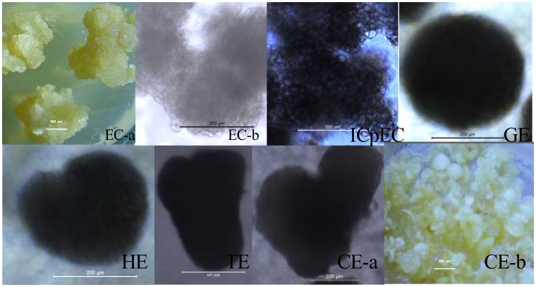
Figure 4. Morphology of embryogenic calli and embryos during the six sequential developmental stages of longan SE.
Developmental stages are indicated at the left of each row. The bars in each phenotypic class are indicated at the middle of each image. The morphology of embryogenic cultures (EC-b, ICpEC, GE, HE, TE, CE-a) were observed using an inverted Leica DMIL LED microscope, except for TE(bar = 500 µm), the bars of others are 200 µm; the images of EC-a and CE-b were both obtained under an Leica DFC295, bars, 600 µm; EC, ICpEC, GE, HE, and TE were cultured on MS medium supplemented with 1 mg/L, 0.5 mg/L, 0.1 mg/L, 0.06 mg/L and 0.03 mg/L 2,4-D, respectively; and the CE was cultured on MS medium.

