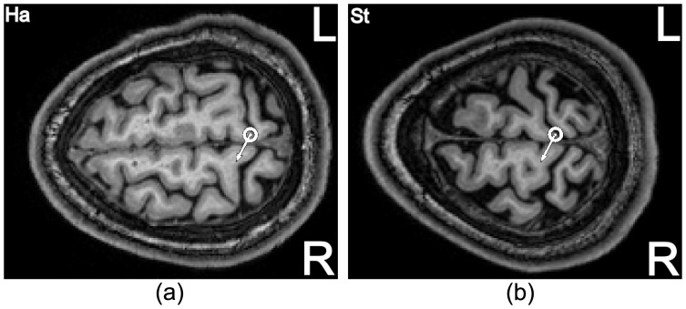
Figure 8. Hot-spot (dot with surrounding circle) for two subjects projected in the MRI images in a transversal view.
An area of the precentral gyrus at the edge to the central sulcus and close to the interhemispheric cleft is in focus for stimulation. The white arrows denote the found optimal coil orientation angle for the individual subject.

