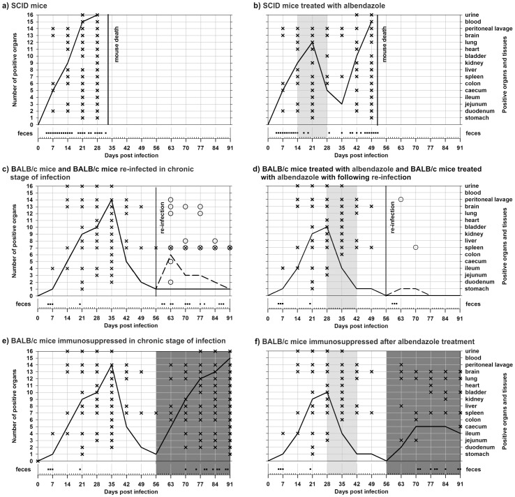
Figure 2. Course of Encepahlitozoon cuniculi genotype II infection, including pattern of spore shedding and dissemination of infection to selected organs and tissues.
a) SCID mice, b) SCID mice treated with albendazole, c) BALB/c mice and BALB/c mice re-infected in chronic stage of infection, d) BALB/c mice treated with albendazole and BALB/c mice treated with albendazole with following re-infection, e) BALB/c mice immunosuppressed in chronic stage of infection, f) BALB/c mice immunosuppressed after albendazole treatment. Light-gray field – albendazole treatment; dark-gray field – dexamethasone immunosuppression; black line – course of E. cuniculi infection; black dash line - course of E. cuniculi re-infection; cross – E. cuniculi positive organ during primarily infection; ring – E. cuniculi positive organ during re-infection; black square – spores shedding during primarily infection; black circle – spores shedding during re-infection.

