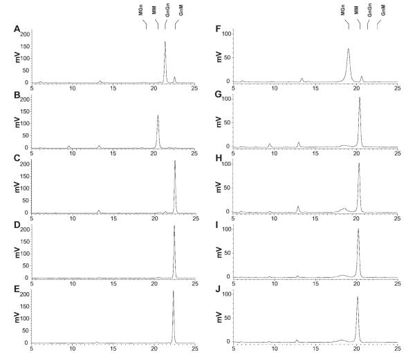
Figure 6. Activity of recombinant insect β-N-acetylglucosaminidases on glycan substrates with an N-acetylglucosamine residue on the α1,3 mannose branch.
Approximately 50 pmoles of the 2-aminopyridine derivatized N-glycan substrates GnGn (left column) and MGn (right column) were incubated for 16 h with solubilized microsomal fractions containing 20 μg of total protein from Sf9 cells infected with wild-type AcMNPV (panels A and F), AcSfGlcNAcase-3 (panels B and G), AcSf-FDL (panels C and H), AcBm-FDL (panels D and I), or AcTn-FDL (panels E and J). The reaction products were then analyzed by reverse phase HPLC with fluorescent detection, as described in Materials and Methods. The markers indicate the elution times of the relevant glycan standards. This reverse phase HPLC method resolves glycans according to the degree of their interaction with the hydrophobic stationary phase. Hence, this method can be used to separate very similar glycans, even if they differ only in the linkages connecting individual sugars. The relative elution positions of the glycans have previously been validated in several studies.17,20,21

