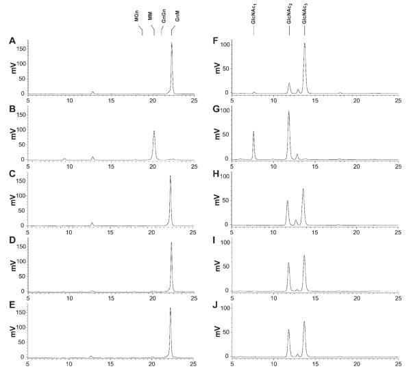
Figure 7. Activity of recombinant insect β-N-acetylglucosaminidases on glycan substrates without an N-acetylglucosamine residue on an α1,3 mannose branch.
Approximately 50 pmoles of the 2-aminopyridine derivatized glycan substrates GnM (left column) and tri-N-acetylchitotriose (right column) were incubated for 16 h with solubilized microsomal fractions containing 20 μg of total protein from Sf9 cells infected with wild-type AcMNPV (panels A and F), AcSfGlcNAcase-3 (panels B and G), AcSf-FDL (panels C and H), AcBm-FDL (panels D and I), or AcTn-FDL (panels E and J). The reaction products were then analyzed by reverse phase HPLC with fluorescent detection, as described under Materials and Methods. The markers indicate the elution times of the relevant glycan standards.

