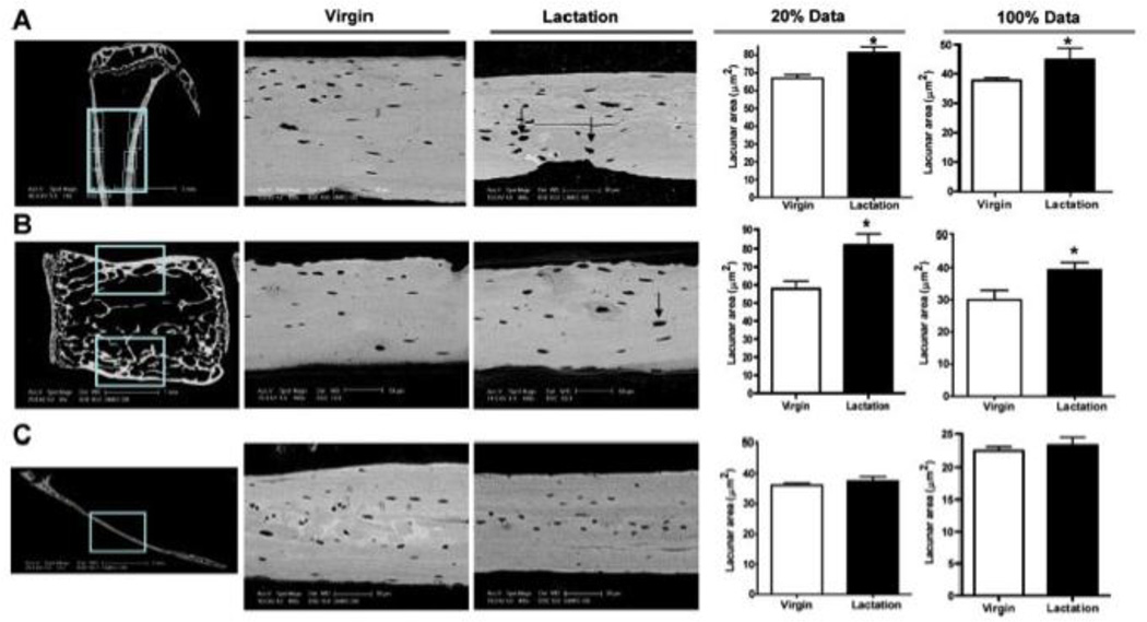
Figure 1. Lactation induces osteocytic lacunar enlargement.
Osteocyte lacunar area as measured by back scatter electron microscopy increases in the tibiae (A) and lumbar vertebrae (B), but not in the calvarium (C). Each panel shows the location of the measurements, representative images and the quantification of the size of the largest 20% of the lacunae and all (100%) of the lacunae in virgin mice as compared to lactating mice. Note that the lacunae are significantly larger in lactating mice in both the tibiae and vertebral bodies, but not in the calvarium. (reproduced from Qing et al, J Bone Miner Res 27:1018–1029, 2012 with permission)

