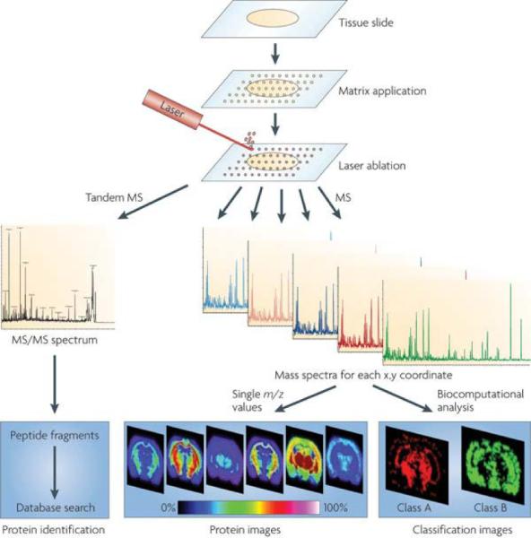
Figure 1. Principle of Imaging Mass Spectrometry.

Schematic of a typical workflow for fresh frozen tissue samples. Sample pretreatment steps include cutting and mounting the tissue section on a conductive target. Matrix is applied to the tissue section and mass spectra are generated at each x,y coordinate for protein analysis or tandem MS spectra for protein identification. Further analytical steps include the visualization of the distribution of a single molecule within the tissue (image) or statistical analysis to visualize classification images as well as database searching to identify the protein. Reprinted with permission from Reference 21. Copyright 2010 Macmillan Publishers Limited.
