Fig 2.
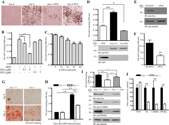
Manipulation of Fyn controls adipogenesis. (A) Inhibition of SFK suppresses adipogenesis. Adipocyte differentiation was induced in 3T3-L1 cells in the presence of DMSO, PP2 (5 μM), or PP3 (5 μM). After 8 days, the cells were fixed and stained with oil red O. Bar, 100 μm. (B) Quantification of stained oil red O in differentiated 3T3-L1 adipocytes treated with different concentrations of PP2 or PP3 (***, P < 0.001; one-way ANOVA; n = 3). (C) Caffeic acid inhibits adipocyte differentiation. Various concentrations of caffeic acid were added to the 3T3-L1 preadipocytes during MDI induction. Eight days after induction, the cells were fixed, stained with oil red O, extracted with isopropanol, and spectrophotometrically quantified (*, P < 0.05; ***, P < 0.001; versus the control; one-way ANOVA; n = 3). (D) Overexpression of Fyn enhances 3T3-L1 differentiation. HA-tagged Fyn CA, Fyn KD, or control vector (GFP) was transfected into the 3T3-L1 preadipocytes. After transfection, the cells were subjected to MDI induction. Eight days after induction, the cells were stained with oil red O and quantified (top panel) (*, P < 0.05; ***, P < 0.001; one-way ANOVA; n = 3). Expressions of the Fyn mutants (middle panel) and GFP (bottom panel) were also verified. (E) Depletion of Fyn in 3T3-L1 cells. 3T3-L1 preadipocytes were transfected with control siRNA (siControl) or siRNA against Fyn (siFyn). After 72 h, expressions of Fyn (upper panel) and tubulin (lower panel) were verified by immunoblotting. (F) Fyn depletion inhibits 3T3-L1 differentiation. Seventy-two hours after siRNA transfection, 3T3-L1 cells were stimulated with the MDI cocktail. Eight days after induction, the cells were fixed, stained with oil red O, and quantified (top panel) (**, P < 0.01; Student's t test; n = 3). (G) Defective adipocyte differentiation in Fyn−/− MEF. MDI inductions were performed in confluent MEF isolated from wild-type or Fyn−/− embryos (E13.5). Eight days after induction, the cells were fixed and stained with oil red O. Bar, 50 μm. (H) Quantification of stained oil red O in differentiated MEF shown in panel G (***, P < 0.001; Student's t test; n = 3). (I) Rescue of adipogenesis in Fyn−/− MEF by overexpressing Fyn. DNA plasmid (pRK5-Fyn) was delivered into Fyn−/− MEF by electroporation. Seventy-two hours after electroporation, the cells were stimulated with MDI cocktail. Eight days after induction, the cells were fixed, stained with oil red O, and quantified (1st panel) (*, P < 0.05; **, P < 0.01; ***, P < 0.001; one-way ANOVA; n = 4). Expressions of Fyn (2nd panel), C/EBPα (3rd panel), PPARγ (4th panel), and tubulin (5th panel) were also examined. (J) Reduced expression of adipogenic markers in Fyn−/− inguinal WAT. Total RNA was isolated from wild-type and Fyn−/− (3-month-old female) WAT and used to perform real-time RT-PCR (**, P < 0.01; Student's t test; n = 3 mice).

