Fig 1.
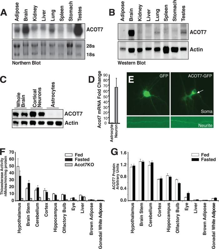
ACOT7 is highly and selectively expressed in neurons. (A) Northern blot tissue profiling of mouse acot7 mRNA. (B) Western blot tissue profiling of mouse ACOT7. (C and D) Western blot (C) or real-time RT-PCR (D) of ACOT7 in cultured rat cortical astrocytes or cortical neurons. (E) Epifluorescence microscopy images of neurites and soma of primary cortical neurons expressing either GFP or ACOT7-GFP. (F and G) Total thioesterase activity (F) or ACOT7 protein abundance (G) measured from total homogenates prepared from fed or overnight-fasted male and female control and Acot7−/− mice; n = 3 to 4. Data represent averages ± standard errors of the means.

