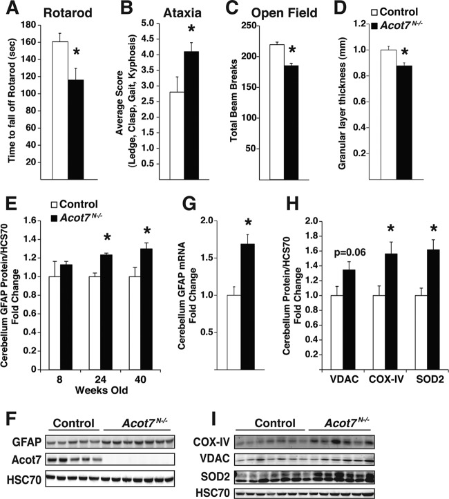
Fig 7.
Loss of ACOT7 results in progressive neurodegeneration and neurological dysfunction. (A and B) Rotarod performance (A) or cerebellar ataxia assessment (B) for 10-month-old control and Acot7N−/− male mice, average of 3 to 5 trials blinded to the observer; n = 6 to 8. (C) Total movement in open field apparatus was monitored over a 40-min period for 3- to 5-month-old male control and Acot7N−/− mice; n = 8 to 10. (D) Cerebellar granular cell layer thickness, as a ratio of lobule width, was measured for Nissl-stained 10-month-old control and Acot7N−/− male mice blinded to the observer; n = 6 to 8. (E and F) Quantification (E) or representative Western blots (F) of GFAP, normalized to HSC70, from male and female control and Acot7N−/− cerebellum of 8-, 24-, and 40-week-old mice; n = 3 to 7. (G) Cerebellum Gfap mRNA abundance, relative to that of Gapdh, from 5- to 6-month-old female control and Acot7N−/− mice; n = 6. (H and I) Quantification (H) or representative Western blots (I) of VDAC, CoxIV, and SOD2 in cerebellum of 24-week-old female control and Acot7N−/− mice; n = 6 to 8. Data represent averages ± standard errors of the means. “∗” indicates P ≤ 0.05 by a two-tail Student t test comparing controls to Acot7N−/− mice.

