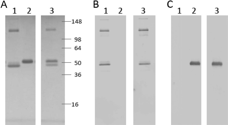
Fig 1.
SDS-PAGE analysis of purified recombinant proteins. After purification from baculovirus-infected cell cultures, antigens were electrophoresed through a 4 to 20% Tris-glycine polyacrylamide gel under reducing conditions and visualized by Coomassie blue staining (A) or Western blot analysis with either an ICP4-specific polyclonal antiserum (B) or a gD-specific monoclonal antibody (C). Prestained molecular mass markers were run on the same gel. Lane 1, ICP4383-766 (300 ng); lane 2, gD2ΔTMR (300 ng); lane 3, ICP4383-766 mixed with gD2ΔTMR (300 ng each).

