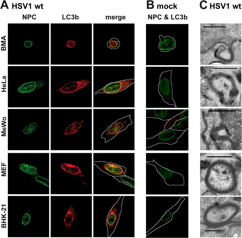
Fig 1.
NEDA is triggered by HSV-1 in different cell types. BMA, HeLa, MeWo, MEF, and BHK-21 cells were infected with HSV-1 wt at a multiplicity of infection (MOI) of 5 for 8 h (A) or mock infected (B), and NEDA onset was analyzed by labeling with antibodies against cleaved LC3b and colocalization with the nuclear pore marker p62 (NPC). (C) HSV-1 wt-infected cells were also analyzed by electron microscopy. Scale bar, 500 nm. White lines denote outlines of cells as observed in transilluminated images in this and the following figures.

