Fig 2.
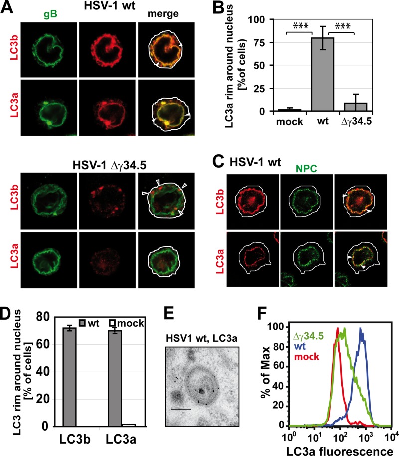
LC3a localizes to NEDA but not macroautophagosomes. (A) Immunofluorescence analysis of BMA cells infected with the HSV-1 wt or Δγ34.5 virus, MOI 5, 8 hpi. Antibodies against both forms of LC3 labeled a perinuclear rim (closed arrowheads) in the presence but not in the absence of γ34.5. The antibody against cleaved LC3b also labeled punctae in the cytosol in the absence of γ34.5 (open arrowheads). (B) LC3a colocalization with the nuclear envelope in mock-infected cells or cells infected with the HSV-1 wt or Δγ34.5 virus. Quantification of three immunofluorescence experiments, 150 cells per condition. Error bars, standard deviations (SD); ***, P < 0.001 in a two-sided Student t test. (C) Immunofluorescence of cells infected with HSV-1 wt, MOI 5, 8 hpi. Nuclear pore complexes (NPC) were labeled with an antibody against p62, and LC3 was detected with isoform-specific antibodies against the cleaved forms of LC3b and LC3a. (D) Quantification of immunofluorescence data with HSV-1 wt-infected and mock-infected cells. About 100 cells for each condition were evaluated; error bars, SD. (E) Immunogold labeling and electron microscopy confirmed the presence of LC3a on four membrane-bound vesicles in HSV-1 wt-infected cells. Scale bar, 250 nm. (F) Flow cytometry analysis of LC3a labeling intensity in BMA cells infected with the HSV-1 wt or Δγ34.5 virus or in uninfected cells.

