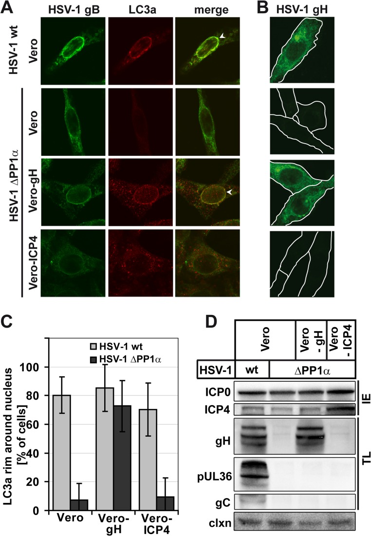
Fig 6.
NEDA is rescued by gH overexpression. Vero cells, Vero CR1 cells that express the late HSV-1 protein gH (Vero-gH), or Vero E5 cells that express the immediate early protein ICP4 (Vero-ICP4) were infected with the HSV-1 wt or HSV-1 ΔPP1α virus at an MOI of 5 for 8 h. (A) Infected cells were detected with an antibody against HSV-1 gB, and NEDA was monitored by LC3a labeling. HSV-1 ΔPP1α caused an accumulation of LC3a around the nucleus (white arrowheads) only in Vero-gH but not wt Vero or Vero-ICP4 cells. (B) Upon HSV-1 ΔPP1α virus infection, Vero-gH cells contained gH levels similar to those of Vero cells infected with HSV-1 wt. (C) Quantification of LC3a nuclear rims in immunofluorescence experiments with the three Vero cell lines in about 50 cells per condition; error bars, SD. (D) Immunoblot analysis of viral protein expression in different Vero cell lines with antibodies against the immediate early (IE) proteins HSV-1 ICP0, ICP4, and the true late (TL) proteins gH, pUL36, and gC. Calnexin (clxn) served as a loading control.

