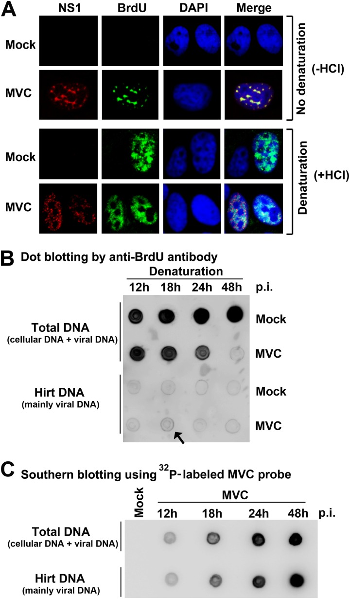
Fig 1.
Cellular DNA replication decreases but still prevails over viral DNA replication during early infection of MVC. (A) Immunofluorescence analysis of DNA replication. WRD cells were seeded on chamber slides 24 h prior to MVC infection. At 18 h p.i., cells were incubated with BrdU for 1 h. The cells on slides were fixed and treated with (+HCl) or without (−HCl) HCl as indicated. Fixed cells were costained with anti-MVC NS1 and anti-BrdU antibodies and DAPI. Confocal images were taken at a magnification of ×100. (B) Dot blot analysis of viral and cellular DNA replication. At the indicated times p.i., mock- or MVC-infected cells were incubated with BrdU for 1 h. BrdU-labeled cells were collected and extracted for total DNA and Hirt DNA (lower-molecular-weight DNA), respectively. The DNA samples were denatured, dot blotted, and immunostained with an anti-BrdU antibody. (C) Southern blot analysis of viral DNA in preparations of total DNA and Hirt DNA. The total DNA and Hirt DNA samples extracted from MVC-infected cells were denatured, dot-blotted, and hybridized with a 32P-labeled MVC NSCap probe (6).

