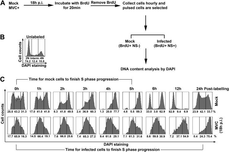
Fig 3.
MVC replication delays host cell S-phase progression. (A) Diagram of BrdU pulsing assay. WRD cells were infected with MVC or mock infected. At 18 h p.i., infected cells were incubated with BrdU for 20 min. After BrdU was removed, cells were taken every hour as indicated in panel C. The cells were treated with HCl and then costained with anti-NS1 and anti-BrdU antibodies and DAPI for flow cytometry analysis. (B and C) DNA content analysis. DNA content was gated as 2N, 4N, and intermediate (Interm.; between 2N and 4N) in unlabeled cells (B) based on DAPI staining, which was used as a reference to gate labeled cells (C) with 2N, 4N, and intermediate DNA content. The numbers under each histogram show percentages of the cell population in each gate.

