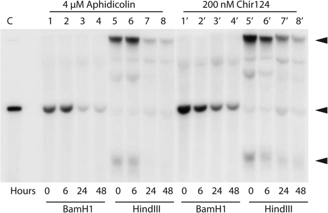
Fig 7.

Southern blot of HPV16 DNA from W12E cells treated with 4 μM aphidicolin (lanes 1 to 8) or 200 nM Chir124 (lanes 1′ to 8′). Prior to loading, the DNA was digested either with BamHI (lanes 1 to 4 and 1′ to 4′), which linearizes the HPV16 episome, or with HindIII (lanes 5 to 8 and 5′ to 8′), which does not cut HPV16. Arrowheads indicate the positions of migration of open circle, linear, and supercoiled HPV forms (from top to bottom) in the uncut samples.
