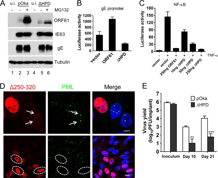
Fig 4.
The contributions of ORF61 HPD to ORF61 functions and VZV virulence in vivo. (A) Melanoma cells infected with pOka or pOka-ORF61ΔHPD were treated with 10 μM MG132 for 6 h. Western blots with ORF61, IE63, gE, and tubulin antibodies are shown. (B) The effect of ORF61 HPD deletion on the gE promoter. Luciferase assays were performed on melanoma cells transfected with the gE promoter and pcDNA constructs expressing ORF61 or the ΔHPD mutant. (C) NF-κB suppression by ORF61 and the ΔHPD mutant. Luciferase assays were performed on melanoma cells that were cotransfected with NF-κB reporter plasmid and increasing amounts (10 to 250 ng) of pcDNA-ORF61ΔHPD and then treated with TNF-α. The graphs show the relative luciferase units (firefly luciferase units normalized with Renilla luciferase units). (D) (Upper panels) Confocal microscopy of melanoma cells infected with the ΔHPD mutant for 6 h and stained for ORF61 (red), PML (green), and nuclei (blue). The images show association of puncta (indicated by white arrows) formed by ΔHPD mutant proteins with PML NBs. Scale bar, 5 μm. (Lower panels) Confocal microscopy of melanoma cells infected with the ΔHPD mutant for 24 h and stained for ORF61 (red), PML (green), and nuclei (blue). The images show PML NB dispersal in pOKa-ORF61ΔHPD-infected cells. Infected nuclei with abundant expression of ΔHPD mutant proteins are outlined by dashed ovals. Scale bar, 10 μm. (E) Replication of pOka and ΔHPD mutant viruses in human skin xenografts in SCID mice. The graph shows the mean titers ± SEM for xenografts that yielded viruses at 10 and 21 days postinfection. **, P < 0.01; ***, P < 0.001; ΔHPD mutant viruses versus pOka (two-way analysis of variance).

