
Fig 2.
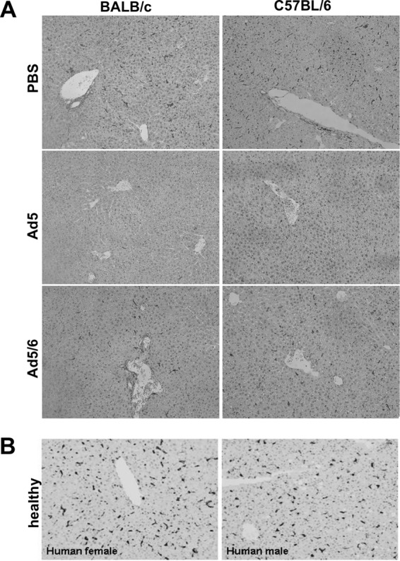
Kupffer cell numbers in mice and humans. (A) BALB/c and C57BL/6 mice were injected with 3 × 10¹⁰ vp of PBS, Ad5GL, or Ad5/6GL expressing GFP-luciferase. After 6 h, the large liver lobe was harvested, formalin fixed, and paraffin embedded. Immunohistochemistry for F4/80+ cells was performed on 4-μm sections. (B) Kupffer cell density in human liver. Normal human liver biopsy tissue was harvested, paraffin embedded, and sectioned (4 μm thick). Immunohistochemical staining for CD68 was used to visualize Kupffer cells.