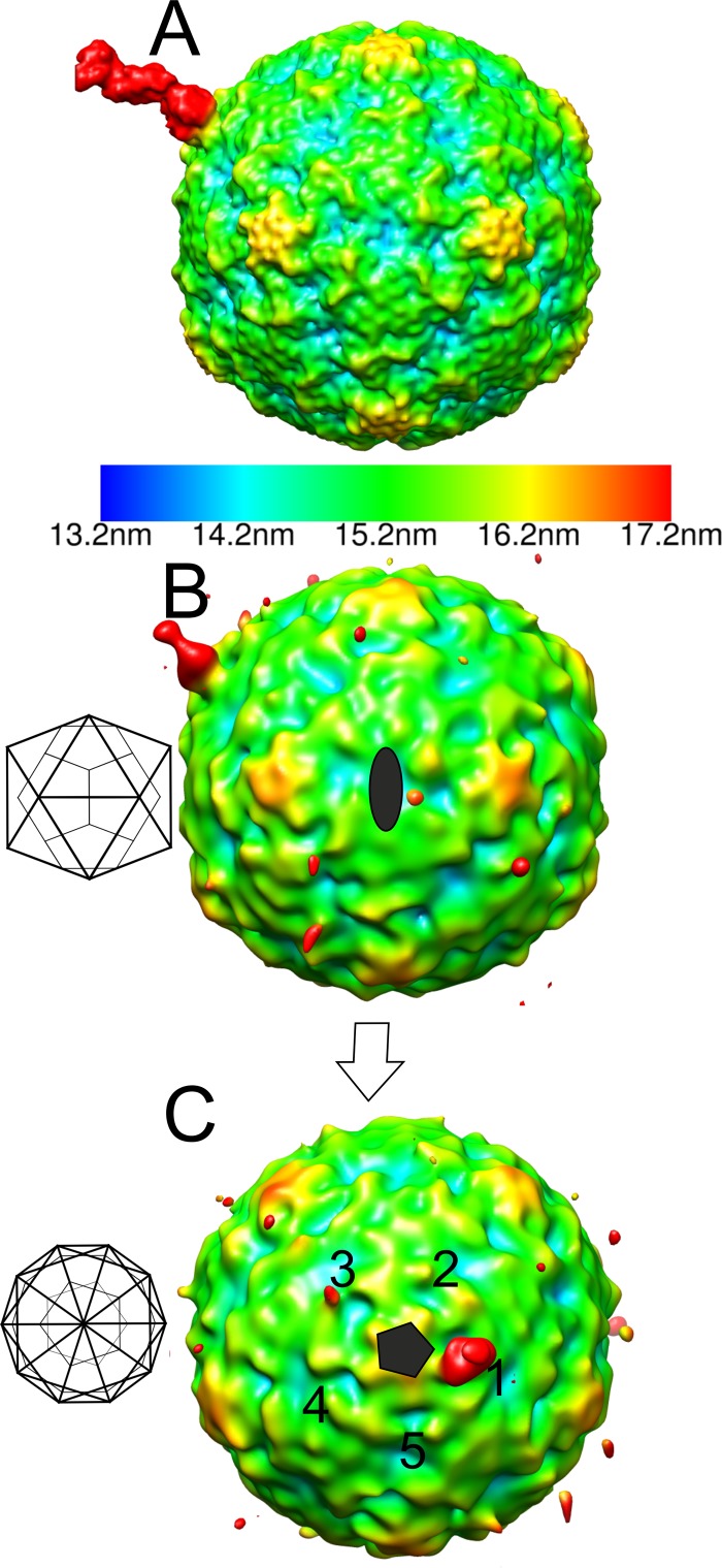
Fig 4.
Radially depth-cued isosurface representations of the asymmetric reconstruction of the filled CVA9-integrin αvβ6 capsid. (A) The reference model used in an asymmetric reconstruction run in AUTO3DEM (41) viewed down a 2-fold axis of symmetry. The reference model has an integrin β-chain in the active state bound to one position adjacent to the C terminus of VP1 on the filled CVA9 capsid reconstruction. (B) The asymmetric reconstruction generated by AUTO3DEM at a 0.5 standard deviation (SD) above the mean, showing the low occupancy of the integrin in the symmetry-related positions on the capsid, where the highest signal is for the position aligned to the modeled integrin shown in panel A. The 2-fold orientation of the capsid is shown as a schematic diagram, which is then rotated to a 5-fold orientation. (C) Five-fold view of the model described for panel B with 5 equivalent positions around one 5-fold vertex marked with numbers 1 to 5. Position 1 is that enforced by the model. Position 3 has a weak signal for the integrin. (A to C) The scale bar for the radial depth cueing is shown in panel A, chosen so that the density at the radii of the integrin is shown in red.

