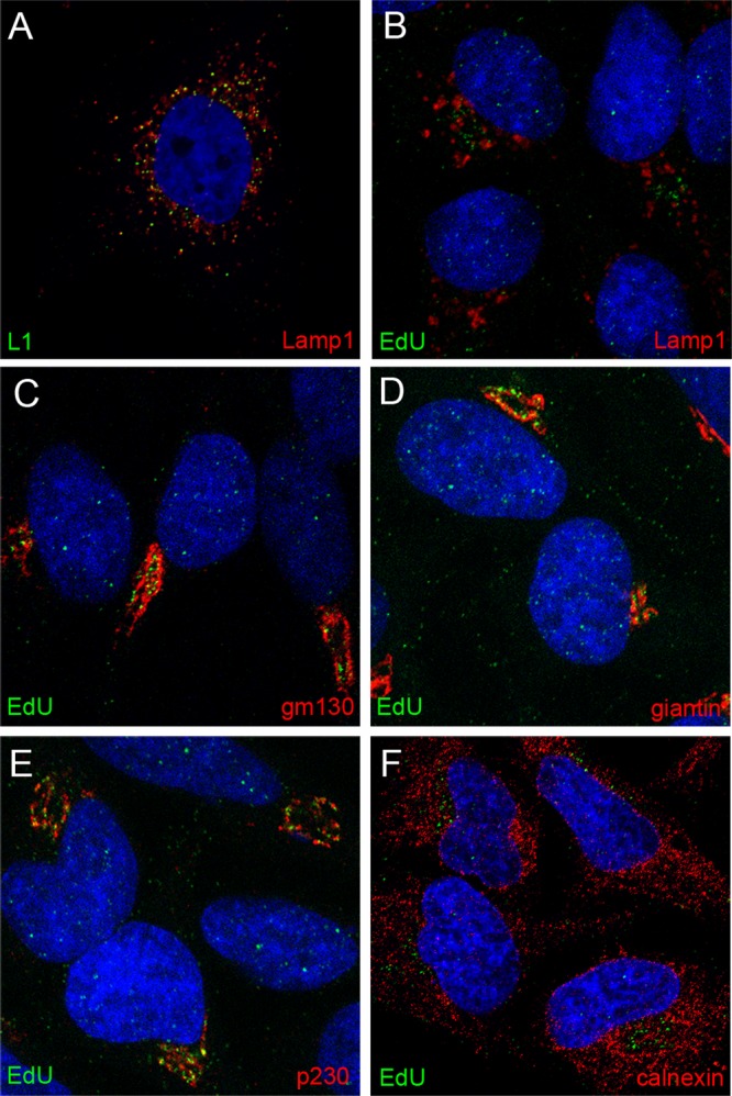
Fig 2.

Localization of pseudogenome relative to subcellular markers. Cells were harvested at 24 h post-virus addition. Reagents used for detection are indicated within the individual panels. Please note that panel A shows HPV capsid staining, whereas all other panels show EdU detection, relative to subcellular markers. The nuclei were detected with DAPI (blue) in all panels.
