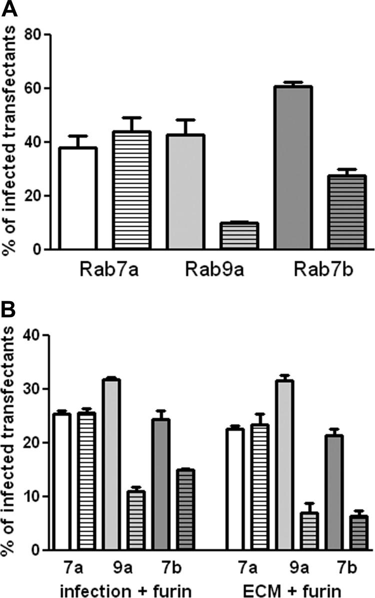
Fig 6.
Effect of expression of DN Rab proteins on HPV16 pseudovirus infection. (A) 293TT cells were transfected with WT or DN Rab expression plasmids. At 24 h, these cells were infected with HPV16 pseudovirus containing a GFP expression plasmid (for Rab7a and Rab9a) or an RFP expression plasmid (for Rab7b). Following 48 h of infection, RFP and GFP coexpression was determined. The percentage of transfected cells that were also infected was determined. The plain bar indicates this percentage for the WT protein; the bar with the horizontal stripes represents the DN protein. Rab 7a is shown in white, Rab 9a in light gray, and Rab 7b in dark gray. (B) The transfected cells were either incubated with exogenous furin during the infection (left side of graph) or plated over furin-cleaved pseudovirus in the presence of furin inhibitor (right side of graph).

