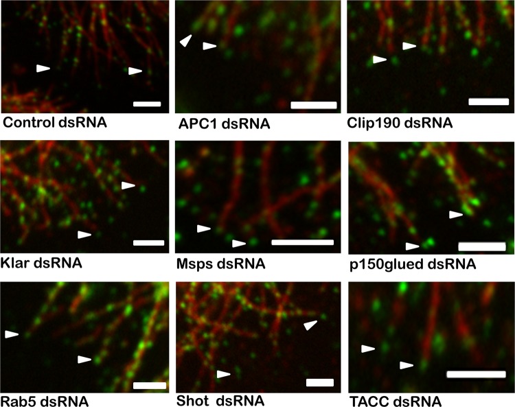
Fig 8.
Localization of CLASP-GFP at the MT plus end. Immunofluorescence for antitubulin and CLASP-GFP in S2R+ cells was used to determine if any of the hits in our ex vivo screen influences the MT +TIP localization of CLASP. The 33 candidate interactors that showed significant MT phenotypes were evaluated for plus-end CLASP localization. Representative images of nine candidates, including the +TIPs and those showing the strongest MT phenotypes, show that CLASP localization to the MT plus end was unchanged for all candidates tested. Bars = 2 μm.

