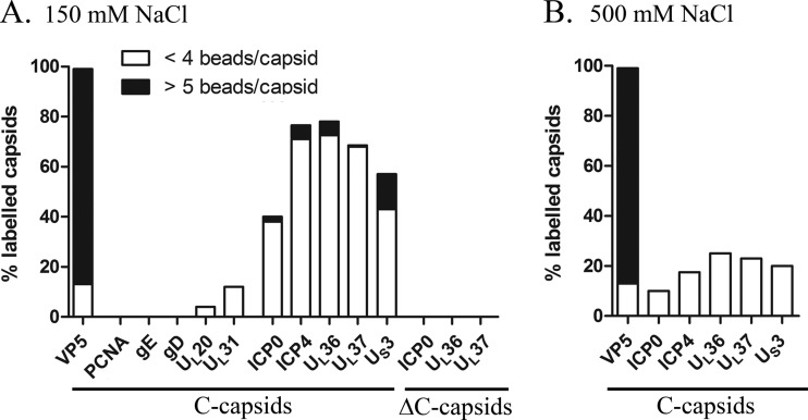
Fig 7.
Immunogold labeling of tegument proteins on nuclear C capsids. (A) Intranuclear wt C capsids were isolated from infected Vero cells on a 150 mM NaCl sucrose gradient and labeled by immunogold with antibodies specific for the following tegument proteins: US3, ICP0, ICP4, UL36, and UL37. Positive-control antibodies included VP5 (capsid control), while negative controls included the viral glycoproteins gD and gE, the viral proteins UL20 and UL31, and the nuclear host marker PCNA. Intranuclear capsids isolated from ICP0, UL36, or UL37 null mutants (ΔC capsids) were also isolated from noncomplementary cells (see Materials and Methods). (B) Intranuclear wt C capsids were isolated from infected Vero cells on a 500 mM NaCl sucrose gradient and labeled by immunogold with antibodies specific for the following tegument proteins: ICP0, ICP4, UL36, UL37, and US3. In panels B and C, quantification of labeled capsids was measured by counting 200 capsids for each sample. Only beads within 10 nm (i.e., one bead equivalent) were considered positive. Strong labeling (black) and weak labeling (white) were determined by the number of beads/capsid. Results are shown as percentages of labeled capsids.

