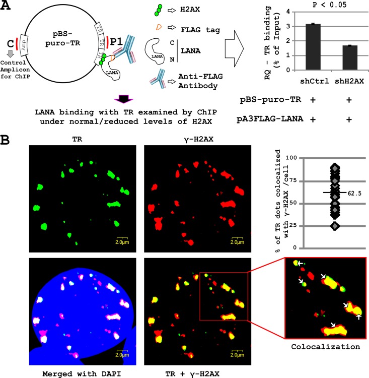
Fig 3.
(A) Role of H2AX in LANA binding with TR. HEK-293 cells were transfected with pA3FL-LANA, pBS-puro-TR, and pGIPz-shCtrl or pGIPz-shH2AX constructs. After 24 h posttransfection, cells were fixed and ChIP was carried out using M2 antibody. Levels of TRs were compared in the different samples. Data have been normalized using an amplicon in the Ampr region of pBS as a reference sequence. (B) Immunofluorescent in situ hybridization (immuno-FISH) was carried out to examine the localization of KSHV TR and γH2AX in HEK-293–BAC36–KSHV cells. Cells were hybridized with the biotinylated KSHV TR probe, followed by incubation with specific antibody against γ-H2AX. The staining was detected by incubation with Alexa Fluor 594 anti-mouse IgG and Alexa Fluor 680 streptavidin conjugate. Alexa Flour 680 staining is shown as a green pseudocolor. Cells were also counterstained with DAPI.

