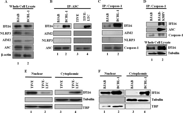
Fig 5.
IFI16 interacts with ASC and caspase-1 and translocates to the cytoplasm in cells latently infected with KSHV. (A) Whole-cell lysates of BJAB and BCBL-1 cells were examined for protein expression with IFI16, AIM2, NLRP3, ASC, and β-actin antibodies. (B) TIVE, TIVE-LTC, BJAB, and BCBL-1 cell lysates were immunoprecipitated with anti-ASC antibody and Western blotted for IFI16, AIM2, and NLRP3 proteins. (C) BJAB and BCBL-1 cell lysates were immunoprecipitated with anti-caspase-1 antibody and Western blotted for IFI16, AIM2, and NLRP3 proteins. ASC and caspase-1 IP blots are shown in Fig. 5. (B and C) (Bottom) IP efficiency. (D) BJAB and BJAB-KSHV cell lysates were immunoprecipitated with anti-caspase-1 antibody and Western blotted for IFI16, ASC, and caspase-1 proteins. Whole-cell lysates were also examined for protein expression with IFI16 and tubulin antibodies. (E and F) Expression and subcellular distribution of IFI16 were examined in nuclear and cytoplasmic fractions of TIVE and TIVE-LTC cells (E) and BJAB and BCBL-1 cells (F) by immunoblot analysis. TBP-1 and tubulin were used to demonstrate the purity and equal loading of nuclear and cytoplasmic fractions, respectively.

