Fig 3.
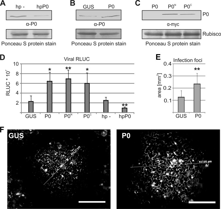
Ribosomal P0 promotes PVA infection. (A) P0 was silenced by expressing an RNA hairpin (hp) that targets P0 transcripts (hpP0), followed by Western blotting using human anti-P autoimmune disease serum directed against the ribosomal P antigen. The empty hairpin vector was expressed as a control (hp −). (B) Exogenous P0 was expressed, and P0 levels were analyzed by Western blotting as described above for panel A. GUS was expressed as a control. (C) Exogenous expression of myc-tagged P0 followed by Western blotting. P0N and P0C have N- and C-terminal myc fusions, respectively. The membrane stained for total protein by Ponceau S is shown at the position of the RUBISCO large subunit as a loading control in panels A to C. (D) Exogenous P0 with or without myc tags or GUS (control) was coexpressed with wt PVA. In parallel, P0 was silenced, and the level of luciferase activity derived from RLUC-tagged wt PVA was determined 7 days after infection (DAI). The Agrobacterium OD600 values used for infiltration were as follows: 0.0005 for wt PVA and 0.5 for P0, GUS, hp −, and hpP0. (E) Fluorescence microscopy of the average area of infection foci formed by GFP-tagged wt PVA at 3 DAI. (F) Representative infection foci from leaves expressing GUS and P0. Scale bar, 200 μm. The number of analyzed foci was >100 for both GUS and P0. *, P < 0.05; **, P < 0.01 (determined by Student's t tests).

