FIGURE 3.
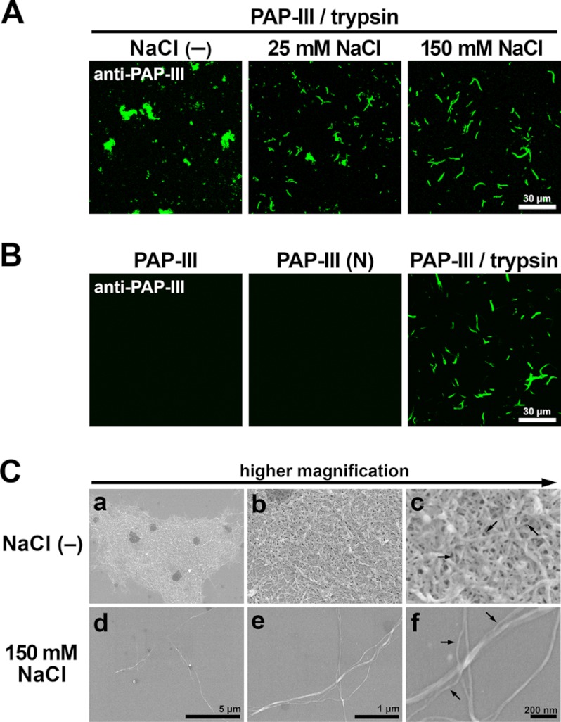
Fibrillar structure of ΔN-PAP-III. A, PAP-III treated with trypsin in 25 mm Tris (pH 7.5) containing 0 (−), 25, and 150 mm NaCl was attached to PDL-coated coverslips and visualized by immunofluorescence using anti-PAP-III antibody. Scale bar, 30 μm. B, full-length PAP-III, PAP-III (N), and PAP-III treated with trypsin in 25 mm Tris (pH 7.5) containing 150 mm NaCl were incubated with PDL-coated coverslips and stained by anti-PAP-III antibody. Scale bar, 30 μm. C, PAP-III treated with trypsin in 25 mm Tris (pH 7.5) containing 0 (−) (a–c) or 150 mm (d–f) NaCl was analyzed by scanning electron microscopy. Scale bars, 5 μm (a and d), 1 μm (b and e), and 200 nm (c and f). Note that filaments with a diameter of 10–20 nm are the minimum constituents in common in both conditions (arrows).

